What Is a Fistula?
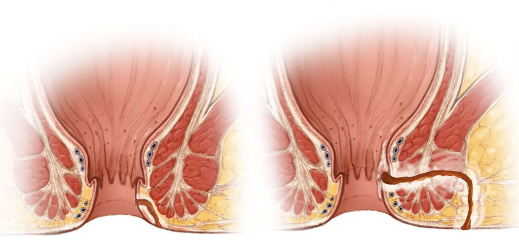
A fistula can be defined as an abnormal connection that occurs between two hollow areas or regions. Fistulas can occur due to various reasons such as an injury, infection, surgery, or inflammation. Although generally due to disease, it can also be created surgically for therapeutic purposes.
Fistulas can occur in any part of the body. Based on the International Statistical Classification of Diseases and Related Health Problems, it can occur in the eye, adnexa, ear, mastoid, circulatory system, respiratory system, digestive system, musculoskeletal system and connective tissue, urogenital system, external causes, or due to congenital malformations, chromosomal abnormalities, and deformations.

1. Types of Fistula
The types of fistula can be categorized based on the patency of the fistula. For example, in a blind fistula, only one end is open while the other end is closed. Blind fistulas can also be known as a sinus tract. In a complete fistula, there are two openings and the fistula is patent.
This means there is both an internal and external opening. In an incomplete fistula, there is one opening on the external side but it does not connect to any internal organ. Although fistulas are generally in the form of a tube, there are also some fistulas that can have multiple branches.

2. Intestinal Fistula
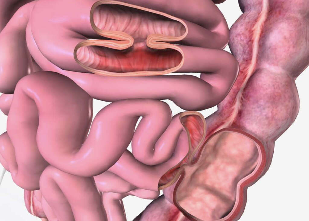
About 75% to 85% of gastrointestinal (GI) fistulas occur due to a surgical complication. The remaining cases evolve spontaneously and can be generally attributed to intraabdominal infection or inflammation.
Fistulas can have a significant impact for patients as it has been shown to increase the mortality and morbidity rates, hospital stays, higher healthcare costs, and delayed return to daily routines. Although the mortality rate has decreased over the decades due to advancement in healthcare (parenteral nutrition and intensive care units), the frequency of intestinal fistulas have remained the same. This has been postulated to be due to increasing complexity of surgical techniques, aging population, and advanced disease.

3. Classification and Risk Factors
GI fistulas can be classified physiologically, etiologically (causes), or anatomically. Anatomical classification can be further divided into external (connecting GI tract and the skin) and internal (connecting GI tract and inside the abdomen).
The GI fistulas can also be classified based on location. Some examples include gastric (stomach) fistulas, small intestine fistulas, colonic fistulas, and aortoenteric fistulas. The possibility of a GI fistula increases in the event of any surgical procedure involving the abdomen, diverticular disease, appendicitis, malignancy (cancer), abdominal trauma, radiation, irritable bowel disease (IBD), GI ulcers, and more.

4. Statistics
In developed countries, the commonest cause of a fistula is Crohn’s disease. It is estimated that as many as 40% of Crohn’s patients develop a fistula. In those who undergo radiation therapy, about 5% to 10% develop a fistula.
In developing countries, obstetric fistulas may be more common due to a higher rate of obstructed labor and lack of early access to emergency care. The outcome of this condition depends on the underlying condition, pain management, wound management, presence of abscess, infection, nutritional status, and more. Patients with an intestinal fistula not only suffer physically, but also emotionally due to the stigma of malodorous fistula drainage. There is also a higher rate of anxiety and depression due to lengthy hospital stays, delay in returning to daily routines, and restriction of social activities.

5. Symptoms and Diagnosis
The symptoms the patient experiences depends on the location of the fistula. Some fistulas are asymptomatic and are only diagnosed as an incidental finding. Some of the symptoms a patient may experience (depending on the location of the fistula) include abdominal pain, diarrhea, weight loss, rectal bleeding, signs of malnutrition, and more.
Some of the lab investigations that can be performed are albumin, prealbumin, blood urea nitrogen, electrolyte, and creatinine levels. A complete blood count can also be beneficial. Abscess culture, urine culture, and urinalysis should also be performed. Procedures such as an endoscopy, fistuloscopy, cystoscopy, or colonoscopy may be indicated in some cases. Medical imaging that may be useful include a computed tomography (CT) scan, magnetic resonance imaging (MRI) scan, ultrasonography, barium enema, and fistulography.

6. Treatment
The treatment of a fistula depends on the location. In the case where an abscess is present, prompt incision and drainage is recommended. Generally, there are several elements that are crucial in the treatment of patients with an intestinal fistula.
This includes proper nutrition, treatment of infection, avoidance of sepsis, and controlling and managing the fistula drainage site. Some GI fistulas can spontaneously resolve without surgery. Conservative management has been described for up to 3 months. When it does not close on its own, some factors to consider include malignancy, presence of foreign body, undrained abscess, and active inflammation. In these cases, surgery may be the best option. Consultations from a nutritionist, wound care specialist, and surgeon may also be beneficial.

7. Hemodialysis Fistula
A hemodialysis fistula is a communication that is surgically created between an artery and vein. Materials such as polyurethane, bovine vessels, saphenous veins, or other synthetic materials can be used as a communication medium between the vein and artery. This is created so it can be routinely used for hemodialysis several times a week.
In the United States, about 430,000 individuals are dependent on hemodialysis. An arteriovenous fistula has been found to be associated with the lowest morbidity, mortality, and cost. Patients with renal (kidney) failure who are not candidates for a renal transplant will be dependent on hemodialysis for the rest of their life. This means having a well-functioning fistula is crucial to provide access for the procedure.

8. Indications and Failures
Throughout a patient’s dependence on hemodialysis, less than 15% of dialysis fistulas remain patent and function without any issues. The average duration after the creation of a fistula is about 3 years while a prosthetic graft will last about 1 to 2 years before there is indication of fistula failure.
With interventions to treat thrombosis and stenosis, the fistula may last about 7 years in the forearm while in the upper arm, 3 to 5 years. Failure and occlusion of the fistula usually occur due to progressive narrowing. Other causes of failure include surgical trauma or obtaining repeated access to obtain blood samples. When in doubt, an interventional radiologist should be consulted.

9. Tracheoesophageal Fistula
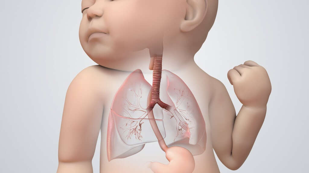
A tracheoesophageal fistula is an abnormal connection that occurs between the trachea and esophagus. It is a common abnormality found at birth but can also occur later in life as a consequence of a surgical procedure.
In a newborn, a tracheoesophageal fistula can cause excessive salivation that is associated with cyanosis, vomiting, choking, and coughing. It can be diagnosed by attempting to insert a nasogastric tube, magnetic resonance imaging (MRI) scan, and gastrografin contrast swallow. A tracheoesophageal fistula can be surgically corrected by resecting the fistula and connecting the segments. Newborns with this issue can have poor feeding and are unable to thrive. They may have other issues such as heart, limb, and kidney deformities.

10. Other Fistulas
A carotid cavernous fistula occurs between the arterial and venous systems in the skull. It can cause progressive visual loss, pain, and bulging of the eye. A labyrinthine fistula is an abnormal opening in the inner ear resulting in leakage of perilymph into the middle ear.
This can cause imbalance, dizziness, and hearing loss. It can occur congenitally or due to a complication of a medical procedure. There are other various fistulas such as lacrimal (eye) fistula, mastoid fistula, coronary arteriovenous fistula, pulmonary vessels arteriovenous fistula, and salivary gland fistula. Generally, a diagnosis can be made via medical imaging. Treatment may be conservative and involve watchful observation or surgery.


