What Is Retinal Detachment?


At the back of our eyes internally is a thin membrane known as the retina. This membrane is one of the key components of our ability to see. Light passes through the eyes and hits the back of the eye where the retina is located. The retina can then detect this light and send the information to the brain via the optic nerve.
Although thin and delicate, the retina is usually quite safe because it is tucked away safely deep in our skull. It is not completely safe, however, as injuries and some medical conditions might cause it harm. One example of this happening is a retinal detachment, and it will cause blindness in some cases.
1. Retinal Tear
A condition that is associated with a retinal detachment is a retinal tear. This can happen because the retina is very thin and cracks in the membrane can develop. In some cases, a retinal tear will appear with no warning and for no apparent reason. In other cases, a retinal tear can be caused by an injury or the vitreous gel pulling on the membrane.
A retinal tear can sometimes result in the eye filling with blood, resulting in a loss of vision. The condition does have the potential to permanently blind the patient in the affected eye, so medical help should be found as soon as possible. A retinal tear can also sometimes cause a retinal detachment.
2. Retinal Detachment
The retina usually lines the inside of the eyeball, fitting quite tightly against the inner surface of the eye. In a retinal detachment, however, the membrane has been pulled away from the inner lining of the eye. This is a condition that is known as a retinal detachment.
A retinal detachment is also a potentially serious condition that has the potential to cause the patient to permanently lose their sight in the affected eye. Getting help from a professional quickly will significantly increase your chances of retaining your eyesight, however. Retinal detachments can be broken down into 3 main types.
3. Rhegmatogenous Retinal Detachments
The most common type of retinal detachment is known as a rhegmatogenous retinal detachment. These are caused by retinal tears because the tear allows fluid to get in between the retina and the interior wall of the eye. The increased pressure from the fluid can then force the retina away from the wall, this resulting in a detachment.
This often happens when the vitreous fluid, which is the jelly-like substance in the eye, shrinks with age. The vitreous fluid will usually just fall away seamlessly when this happens. However, it can sometimes pull on the retina, and this can result in the retina becoming torn.
4. Exudative Retinal Detachments
This type of retinal detachment is not caused by a tear in the retina. Instead, it is caused by other factors that can result in fluid building up between the retina and the internal wall of the again. As with rhegmatogenous retinal detachments, this build-up of fluid can cause the retina to be forced away from the eye’s interior wall.
Reasons why this can happen include inflammatory disorders of the eye, or tumors. Injury to the eye is another potential cause of exudative retinal detachments. Macular degeneration is another potential factor, which is the degradation of the central part of the retina. This is something that tends to happen as people get older.
5. Tractional Retinal Detachments
In some people, scar tissue can develop on the retina. When this happens, the retina can be pulled away from the eye as the scar tissue tightens the retinal tissue. This can result in a variety of retinal detachment that is known as a tractional retinal detachment.
This will sometimes happen in people who have sustained damage to the blood vessels located in the retina. It is a relatively common condition in people who have diabetes that is left uncontrolled. Regardless, the condition should be treated as soon as possible to help ensure the patient does not lose their eyesight.
6. Symptoms
A retinal detachment will not cause any discomfort for the patient. However, other symptoms will likely appear that will let the patient know that something is wrong. This includes a phenomenon known as photopsia, which means that patient is seeing flashes of light in their vision.
A retinal detachment will also often cause the patient to see many of what are known as floaters. These are the appearance of fiber-like structures in your vision that you are unable to focus on. Other potential symptoms include a reduction in the patient’s peripheral vision, and blurred vision. These symptoms should encourage the patient to speak with a professional, especially if they develop suddenly.
7. Who’s At Risk
Anybody can experience a retinal detachment, but some people are more prone to the condition than others are. One of the main factors is age, and people above the age of 50 are at an increased risk. People who are near sighted are also more prone to developing a retinal detachment.
You are also more likely to experience a retinal detachment if there is a history of the condition in your family. Having experienced an eye injury at some point in the past will also increase the risk. Another potential factor is diseases of the eye, having had eye surgery in the past, and a previous retinal detachment.
8. Diagnosis
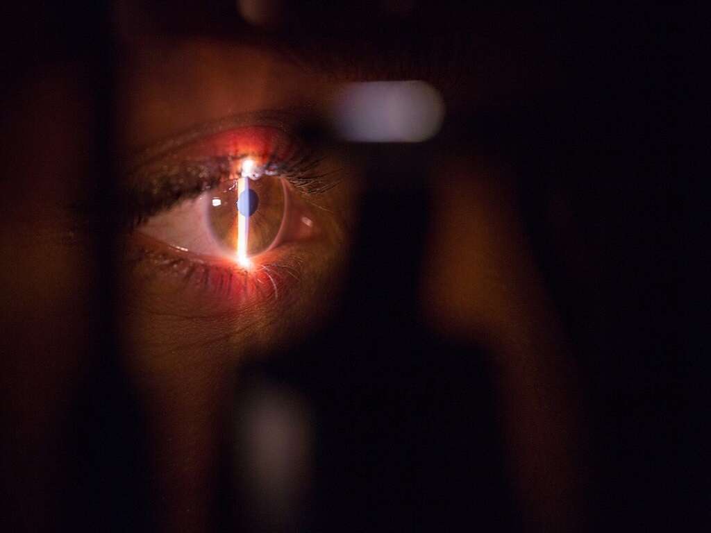
In order to be able to confirm the cause of your symptoms, the optometrist will need to exam your eyes using specialized tools and equipment. This will include ultrasound technology that uses sound waves to help see if there is any bleeding inside the eye.
A retinal examination may also be used, which uses a specialized lens to look at the back of the eye. This will allow the experts to visually see whether or not there is a tear or detachment in the retina or any other problems. If a tear or detachment is not detected, you may be asked to return again in a few weeks.
9. Retinal Tear Treatment
Potential treatment for a retinal tear includes cryopexy. This involves applying a freezing element to the outer surface of the eye. This is performed on the part of the eye that is immediately above the location of the tear. This will result in a scare, and this scar will help fasten the retina to the eye wall. The procedure is performed under a local anaesthetic.
Another possible treatment is photocoagulation. This procedure also involves creating a scar to help fasten the retina to the eye wall. This time, however, a laser beam is used instead of freezing the affected area. The patient should take it easy for 2 weeks to help allow the eye to heal properly.
10. Retinal Detachment
Potential treatments for a retinal detachment include a vitrectomy. In this procedure, the vitreous fluid is removed from the eye. It is then replaced with another substance that will fill the eye, causing the retina to be pushed back against the eye wall. Scleral buckling is another potential treatment.
Scleral buckling involves causing an indent in the eye, pushing the eye wall closer to the retina. There is also pneumatic retinopexy, and this involves injecting gas or air into the patient’s eye. This will help to push the area back against the eye wall. Cryopexy is also often used in conjunction with this procedure.