What Is Pleurisy?


To take in fresh air, our muscles will pull on our lungs to open them up, thus sucking air in. The lungs are then pushed upon to cause them to deflate, and this will cause us to exhale. This is an effective system of ensuring our bodies have a constant supply of fresh air from which they can take oxygen.
As the lungs expand and contract, they could rub against the inside of the chest cavity, and this would cause problems. However, we have evolved a system that allows the two to slide smoothly against each other. Things will sometimes go wrong with this system, however, including a condition known as pleurisy.
1. Pleurisy
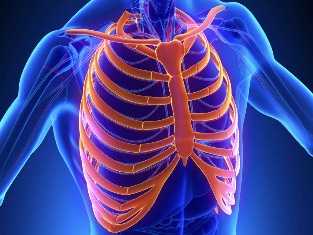
In the chest cavity are two layers of tissue known as the pleura. One of these lines the inner wall of the chest, and the other lines the outer wall of the lungs. The two layers are right next to each other, except for being separated by a small gap that is filled with a fluid.
The two layers allow for the lungs to slide over the inner wall of the chest as we inhale and exhale. Pleurisy is a condition where these layers become inflamed, and this will cause the two layers of the pleura to rub against each other. This will, in turn, result in some very unwelcome symptoms for the patient.
2. Infection
Among the most common causes of pleurisy is an infection of sorts. This can mean viral infections and pleurisy will also sometimes be caused by fungal infections, or bacterial infections. Among the most common type of infection that causes pleurisy is pneumonia, which is an infection of the interior lining of the lungs.
Pneumonia will mean that the patient has difficulty exchanging gases in their lungs in what has the potential to be a very serious condition. Another potential cause of pleurisy is tuberculosis, which is another type of bacterial infection. Like pneumonia, tuberculosis can be treated but is also potentially very dangerous.
3. Injuries
Pleurisy is also sometimes caused by physical injuries to the chest area, including fractures of the ribs. Such injuries will include those sustained in road traffic accidents and similar. Pulmonary embolisms are other potential causes of pleurisy, which is caused by a type of blood clot.
A pulmonary embolism is a type of blood clot that formed elsewhere in the body. These clots can become dislodged and travel through the blood stream until they eventually become stuck elsewhere. Blood will then not be able to pass through the blocked blood vessel causing what should be treated as an immediate medical emergency.
4. Autoimmune Disorders
Autoimmune disorders are conditions where the patient’s immune system is attacking certain parts of their body. Examples of such disorders that can result in pleurisy include lupus and rheumatoid arthritis. Such conditions tend to be chronic, although they can also usually be managed to a degree.
Any lung cancer that is located near the pleura can also cause pleurisy. Some diseases like sickle cell disease are another potential cause of the condition. Sickle cell disease is an abnormality with red blood cells which can cause symptoms that also include pneumonia. Certain types of medication will also be the cause of pleurisy in some cases.
5. Symptoms
The symptoms of pleurisy typically include a pain in the chest that gets worse when the patient coughs or sneezes. Even breathing will be enough to cause the patient pain in some cases. This can cause the patient to try and breathe shallowly to avoid pain, while their lungs might automatically stop expanding in response to the pain.
Depending on the cause of the condition, the patient may also have a fever. This tends to be the case if the pleurisy is being caused by an underlying infection. Pleurisy can also sometimes cause a cough, and this can aggravate some of the other symptoms further.
6. Pleural Effusion
As mentioned, there is usually a small amount of fluid between the layers of pleura, and this helps to act as a lubricant. Pleural effusion is a condition where more than usual volumes of fluid accumulate between the two layers. The condition will sometimes be present at the same time as pleurisy.
Symptoms of pleural effusion include chest pain, shortness of breath, cough, and a fever. Although it can have negative symptoms, pleural effusion will sometimes help reduce the symptoms of pleurisy by providing more of a buffer between the layers of pleura. The fluid will sometimes be drained, although doctors will usually try and treat the underlying cause first.
7. Empyema
In some cases, the additional fluid in between the pleura can become infected, and this can cause pus to accumulate in the pleural space. There are numerous reasons this can happen, and recently having had pneumonia is among the most common. Some surgical procedures can also cause the space to become infected.
Typical symptoms of empyema include a dry cough and being short of breath. A pain in the chest is another symptom, and empyema will also sometimes cause headaches. The condition can also cause a loss of appetite, confusion, and excessive sweating. The pus will need to be drained from the cavity to help prevent severe complications.
8. Atelectasis
Excess fluid in the pleural space is not usually too much of a problem for the patient. However, if there is a significant volume of fluid then it can cause pressure to build up. If there is enough pressure then it can cause a partial or complete collapse of a lung in what is known as atelectasis.
Atelectasis will often not cause any clear symptoms. When there are symptoms, however, they will typically include difficulty breathing. The patient may also be wheezing and they may also be breathing shallowly and more rapidly than usual. Atelectasis will also sometimes cause a cough.
9. Diagnosis
Your doctor will need to perform a physical exam. This will likely include the use of a stethoscope which will allow your doctor to listen inside the chest cavity. They will also need to ask about your symptoms and about your medical history. A blood sample will likely be taken to help experts to look for signs of an infection.
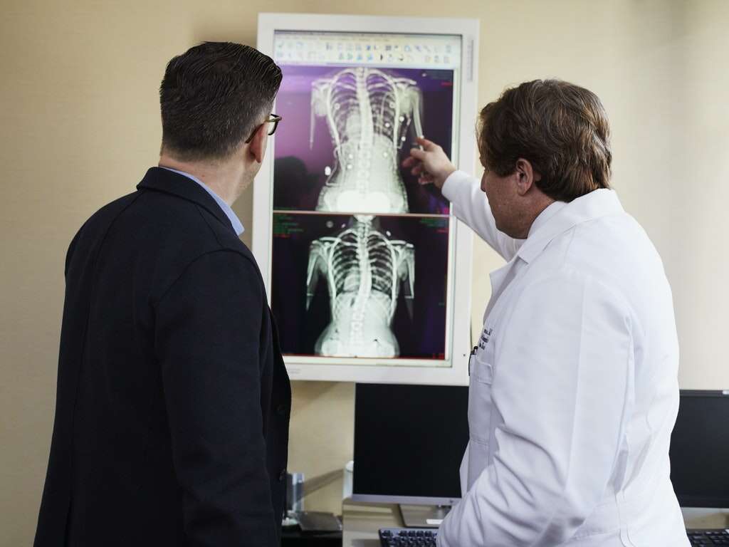
Imaging tests include CT scans, X-rays, and ultrasounds will also be used, and these will help experts look for physical signs of pleurisy. An electrocardiogram may also be requested to help experts to measure electrical activity in the heart. This will help to rule out heart conditions that might causing the pain.
10. Treatment
The treatment for pleurisy will depend largely on what is causing it. If it is a viral infection causing it then the patient will usually be asked to rest and let the immune system deal with it. If it is caused by a bacterial infection, however, then the patient will likely be given an antibiotic to help clear up the infection.
Other medication may also be prescribed to help treat the patient’s symptoms, and the patient should stop smoking at least until the condition clears up. Associated conditions may also need to be treated, and this includes draining excess fluid from the pleural space.