What Is Meiosis?


All species on Earth reproduce to help continue the species, but some have some very different methods of procreation than others. In human beings, copulation occurs that enables the male sperm cells to be introduced to female ovum (eggs). This method allows DNA to be shared by both parents, allowing each of their characteristics to be adopted by their children.
In order for this to happen, suitable cells need to be produced to enable procreation. This is achieved in humans by a process known as meiosis. Here’s a closer look at what meiosis is and the various stages that are involved.
1. Interphase
The first stage in the meiosis process is known as the interphase. It is also the first step in Meiosis I. The first step is for the DNA in the cell to be copied, resulting in two identical sets of full chromosomes. The DNA is basically the blueprint that determines much about us physically and mentally.
In addition, there are two centrosomes in the cell. These consist of two microtubule rings known as centrioles. The purpose of these rings is to help provide structure for the cell by organizing microtubules. They are also responsible for helping to pull apart chromatids during the cell division process.
2. Prophase I
The next step in meiosis is prophase I. The two separate and identical strands of DNA now come together and form x-shaped structures. The shapes of these structures are clearly visible under a reasonably powerful microscope.
Each of the chromosomes is made of two chromatids, which is the name given to the thread-like strands that are divided longitudinally. The two chromosomes then match up accordingly and exchange parts of DNA. This latter process is known as crossing over and/or recombination. The membrane around the nucleus will then dissolve, allowing for the release of the two chromosomes, and the meiotic spindle is extended across the cell.
3. Metaphase I
The next stage in the process is known as the metaphase I. In this stage, the pairs of chromosomes line up with each other at the center of the cell, which is also known as the equator of the cell. At this stage of the process, the centrioles are now on opposite sides of the cell to each other.
At this point, the centrioles will also have the meiotic spindles extending outward from each one. The fibers of the meiotic spindles also attach to each of the pairs of chromosomes. This allows them to take a grip on the chromosomes in preparation for the next phase.
4. Anaphase I
Anaphase I is the name that is given to the next step in the process. As already mentioned, the fibers of the meiotic spindle have already attached themselves firmly to each of the chromosomes.
The meiotic spindles will then pull on each of the chromosome pairs. This will pull the pair of chromosomes away from each other breaking the link in the center of each. We are now left with two separate chromosomes, each with chromatids that were shared from each other. This latter part of the process is what differentiates meiosis from mitosis, which is the process responsible for reproducing other types of cell.
5. Cytokinesis and Telophase I
Next up in the process is cytokinesis and telophase I. Now, it becomes visible under a microscope that the cell is beginning to “pinch” together in the middle. This is in preparation for the cell to eventually separate into two separate cells completely.
By now, each of the chromosomes has made their move toward each of the centrosomes. The full set of chromosomes are now gathered at each pole of the cells close to the centrioles. A membrane will now form around each set of chromosomes. This latter process, known technically as cytokinesis, helps to create two separate nuclei.
6. Prophase II
The next phase is prophase II, which is also the first phase in Meiosis II. At this stage of the process, there are now 2 separate cells, which are known as daughter cells. Each of these cells now has 23 chromosomes, which themselves have 23 pairs of chromatids. In each of these daughter cells, the chromosomes again condense into structures that are x-shaped.
After the split, each new cell was left with only one centriole again, but they now duplicate to make two centrioles in each of the daughter cells. In addition, the meiotic spindle forms again as it did in the metaphase I phase.
7. Metaphase II
Next, we have the metaphase II stage of the meiosis process. Here, the existing x-shaped chromosome structures will line up at the cells equator.
In addition, the centrioles will line up at opposite poles of each of the cells, as was the case in the prophase I stage we described earlier. As mentioned, the meiotic spindle has formed again, and the spindles fibers will attach firmly to each of the chromosome structures in the same manner that they did in the metaphase I stage. This helps to prepare each of the structures for the next stage of the meiosis process.
8. Anaphase II
Anaphase II is the next step in the process. As already mentioned, fibers from the meiotic spindle have already attached themselves firmly to the chromosome structures. They will now pull the structures apart.
This process is different from the separation that takes place during the anaphase I stage. Instead of ending up with two separate chromosomes with one in each cell, we now four separate chromatids, with two in each cell. The chromatids are now also chromosomes in their own right. They are now known technically as unreplicated chromosomes. This stage is important because it helps to ensure that there are equal sets of chromatids in each cell.
9. Cytokinesis and Telophase II
Next we have the cytokinesis and telophase II stage. In this stage, each of the chromosomes will move toward the poles of each cell, pulled into position by the fibers of the meiotic spindles. This is in preparation for the splitting of the cells because it ensures that we are left with one set of chromosomes in each cell.
Cytokinesis then occurs, which is the process of the cells physically separating from each other. We are now left with four separate cells which, at this stage, are known as granddaughter cells. These cells are basically the end of the process and are almost ready for fertilization.
10. Granddaughter Cells
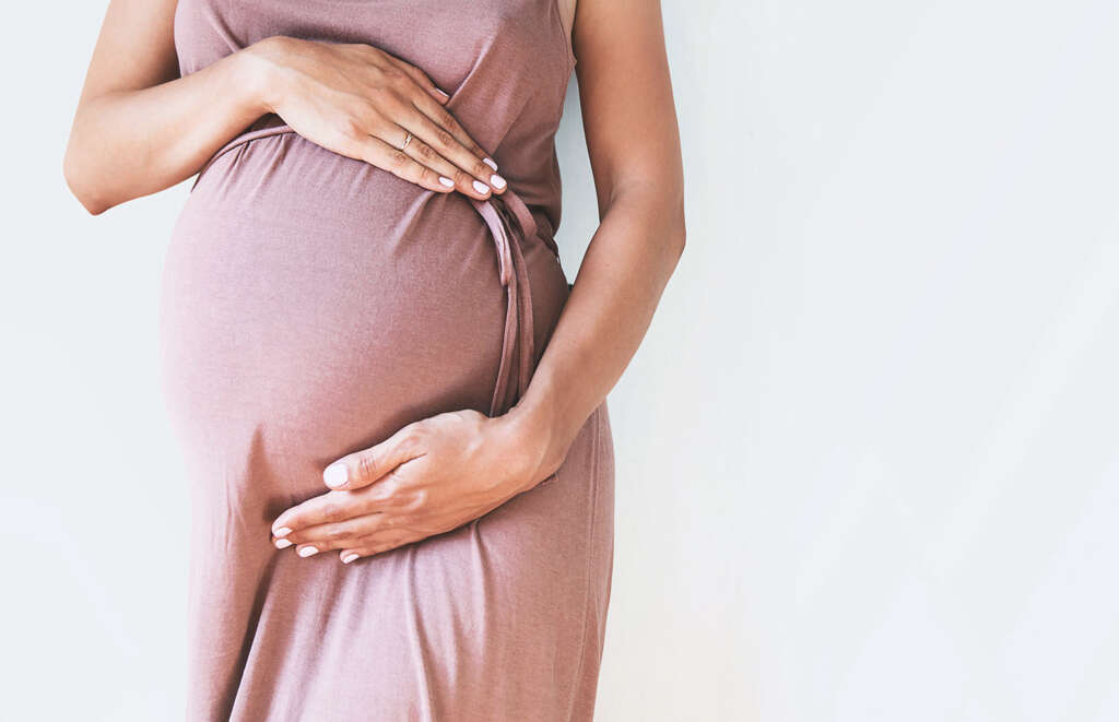
As mentioned, we are now left with four individual granddaughter cells. These cells are haploid, and this means that they have half the number of chromosomes that a parent cell does. These cells are essential for the reproduction process. In women, granddaughter cells are basically eggs and, in men, the cells are sperm cells.
After copulation, a sperm cell will often find the egg and the two cells will share their DNA, and the egg will become fertilized. The fertilized egg, now known as a zygote, will share DNA from both the male and female parents. It will begin to develop further after settling in the womb.