What Is Mastocytosis?
Mastocytosis is a condition where there is proliferation and accumulation of mast cells in organs such as the skin. According to the World Health Organization (WHO), mastocytosis can be classified into indolent systemic mastocytosis, cutaneous mastocytosis, aggressive systemic mastocytosis, mast cell sarcoma, mast cell leukemia, extracutaneous mastocytoma, and systemic mastocytosis with associated hematologic non-mast cell lineage.
Since cutaneous mastocytosis is the commonest form of the disease, this article will be focusing on cutaneous mastocytosis. Cutaneous mastocytosis can be further divided into diffuse erythrodermic mastocytosis, solitary mastocytoma, urticaria pigmentosa, and paucicellular mastocytosis. Among these forms, urticaria pigmentosa is the commonest form. Mastocytosis is a rare disorder that affects adults and children due to the accumulation of functionally defective mast cells.

1. Pathophysiology
Mastocytosis is now categorized among the myeloproliferative neoplasms. In cutaneous mastocytosis, the increased concentrations of soluble mast cell growth factor stimulates melanin pigment production, mast cell proliferation, and melanocyte proliferation. This mechanism also explains the darkening of skin often seen in cutaneous mast cell lesions while the itching has been thought to be due to the production of interleukin 31.
Since interleukin 6 levels are elevated in mastocytosis, it indicates that it may be involved in pathophysiology of mastocytosis. Associated symptoms are thought to be due to the release of mast cell derived mediators such as acid hydrolases, neutral proteases, heparin, prostaglandins, and histamine.
2. Epidemiology
Based on the number of new patients who visit dermatology clinics, it is estimated that 0.1% to 0.8% have some type of mastocytosis. In children, the commonest type is maculopapular cutaneous mastocytosis at about 47% to 75% of cases. Other types in the pediatric population include mastocytoma and diffuse cutaneous mastocytoma.
Mastocytosis is most commonly seen among children where 75% occur in infancy or early childhood. The incidence of mastocytosis peaks again in the population between the ages of 30 to 49 years. Most mastocytosis cases are seen among whites as the cutaneous lesions are less obvious in individuals with heavily pigmented skin.

3. Signs and Symptoms
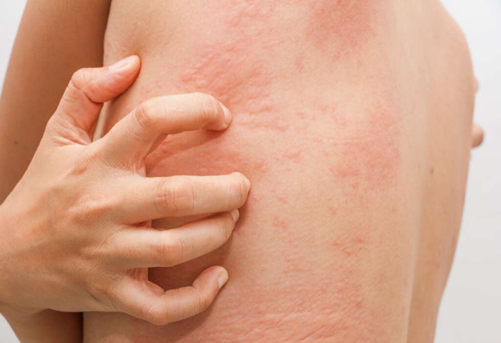
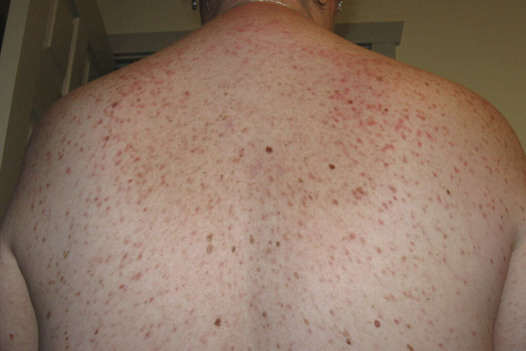
Patients with mastocytosis can present with cutaneous lesions that are hyperpigmented (yellowish to reddish brown) and itchy. The skin lesions may appear as nodules, macules, papules, bullae, or blisters. In some cases, patients with extensive disease tend to experience acute symptoms that are aggravated by the ingestion of specific foods, drugs, and activities.
Some of the symptoms include dyspnea, headache, flushing, rhinorrhea, wheezing, diarrhea, vomiting, nausea, and syncope. Systemic involvement may manifest as a new fracture and bone pain as chronic exposure to stem cell factor and heparin increases the risk of osteoporosis. Other associated symptoms include abdominal cramps, weight loss, irritability, malaise, and chest pain.

4. Diagnosis
In urticaria pigmentosa, diagnosis can usually be achieved via the history and examination of the patient. However, as a confirmatory test, a skin biopsy may be necessary using a 3 mm to 5 mm punch biopsy. If systemic disease is suspected, the serum tryptase levels can be measured.
To diagnose systemic mastocytosis, the criteria (one major and one minor or 3 minor) has to be fulfilled. The major criterion is the presence of dense infiltrates of more than 15 mast cells in an extracutaneous organ or bone marrow. Minor criteria include the finding of mutation in KIT (D816V), aberrant mast cell morphology, aberrant phenotype on the mast cells, and S-tryptase levels of more than 20 ng/ml. Imaging tests such as a bone scan, radiologic survey, computed tomography (CT) scan, and endoscopy may also be beneficial.
5. Medication
Treatment for mastocytosis tends to be conservative and supportive. It includes symptomatic relief. Patients should avoid triggers and medications that precipitate the release of mediators.
Medication includes the use of H1 and H2 antihistamines, oral disodium cromoglycate, cautious administration of aspirin, leukotriene antagonists, mast cell stabilizers, proton pump inhibitors, salbutamol, epinephrine, corticosteroids, and antidepressants. In advanced disease, cytoreductive therapy such as alpha interferon, cladribine, and tyrosine kinase inhibitors may be beneficial as well.

6. Further Management
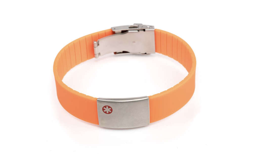
As previously mentioned, patients should wear medical alert bracelets and a self injectors should be prescribed. General anesthesia may cause issues in patients with systemic mastocytosis. Due to the complexity of treatment for systemic mastocytosis, a hematologist may be the best option for optimal management of the disease.
In cutaneous mast cell lesions, some studies have reported cosmetic improvement when treated with the 585-nm flashlamp pumped dye laser and 532-nm Nd:YAG laser. Hospitalization and emergency resuscitation may be necessary for those experiencing hypotensive shock or syncope due to sudden severe degranulation of the mast cells. In aggressive systemic mastocytosis, allogeneic stem cell transplantation may be an option.
7. Consultations
In some cases of mastocytosis, a consultation may be required for staging and bone marrow biopsy. As per the WHO classification of systemic mastocytosis, several staging investigations help define the disease subtype.
When the serum tryptase level is more than 200 ng/ml or there is more than 30% bone marrow mastocystosis burden, it is indicative of a high systemic mastocytosis burden. Hemoglobin levels less than 10 g/L, absolute neutrophil count less than 1000 g/L, platelet count less than 100,000 g/L, palpable spleen due to enlargement with hypersplenism, liver enlargement with ascites and impaired liver function, malabsorption with low albumin and weight loss, and osteolysis or severe osteoporosis can result in life-threatening organopathy or pathologic fractures.

8. Diet and Activity
Some doctors may advise patients with cutaneous mastocytosis to avoid foods and beverages that can trigger the release of mast cell mediators such as spicy foods, hot beverages, crawfish, lobster, salicylates, alcohol, and cheese.
Although there is some evidence that has shown alcohol can be a trigger, the other foods causing a reaction remain hypothetical and require further research. Activity-wise, patients with mastocytosis should avoid physical stimuli such as physical exertion, extreme temperatures, emotional stress, bacterial toxins, and insect bites to which the individual is allergic to as the condition can be worsened by rubbing and scratching of the cutaneous lesions.

9. Prognosis
The outcome depends on the age of onset of mastocytosis. In urticaria pigmentosa where most patients exhibit symptoms before the age of 2 years old, there is an excellent prognosis and the condition tends to resolve by puberty. It is estimated that the lesions diminish by 10% every year. Life threatening episodes of shock are rarely caused by acute extensive degranulation. Serum baseline tryptase levels can be used to predict the severity of the disease.
Those with adolescent or adult onset urticaria pigmentosa have a poorer outcome as the condition is more likely to be persistent with a higher risk of systemic involvement. The malignant transformation rate for juvenile onset systemic mastocytosis is 7% while in adults it increases to as high as 30%. After the age of 10 years, cutaneous mastocytosis has a poorer prognosis with more likelihood to be persistent, systemic disease, and malignant transformation.

10. Patient Education and Research
Patients with cutaneous mastocytosis should try to avoid substances and physical stimuli that triggers the condition. Awareness regarding the signs, symptoms, and treatment of anaphylaxis is also important especially among those with severe symptoms or systemic disease.
It is recommended that those with systemic disease or severe symptoms to wear a medical alert bracelet, inform teachers at school or colleagues at work, and carry injectable epinephrine in case of an acute event. Research has improved the diagnosis of mastocytosis and the identification of factors and genes that causes increased mast cell production.
