What Causes Moles?
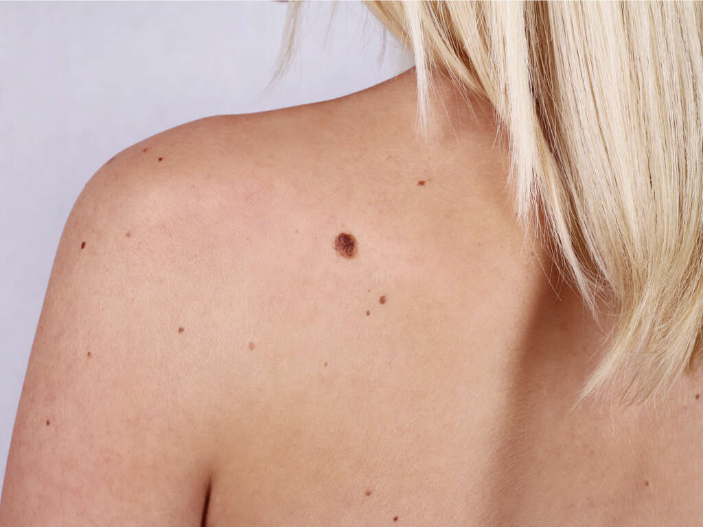
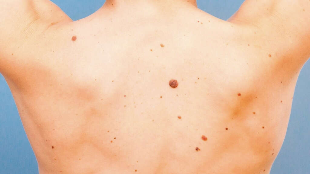
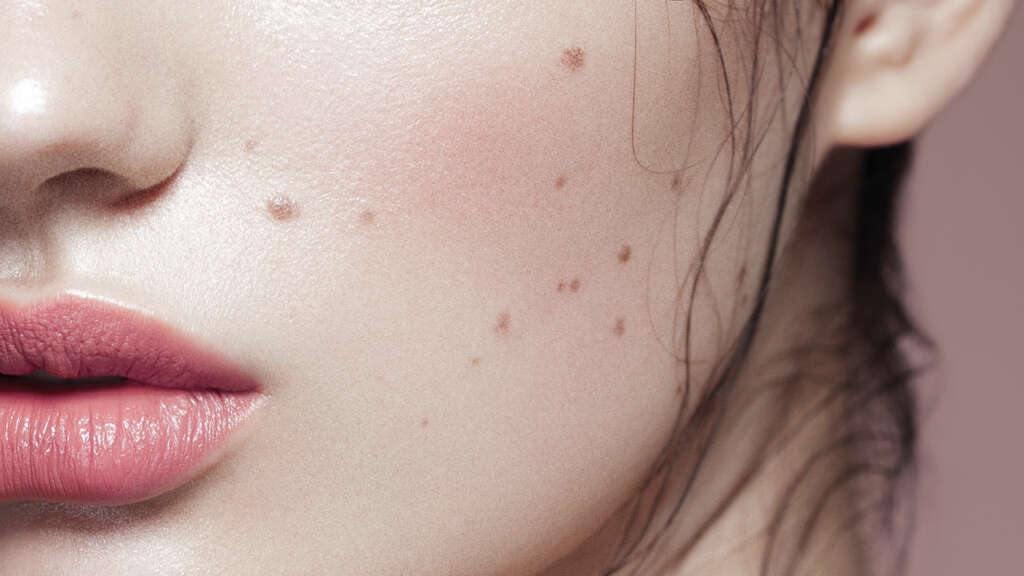
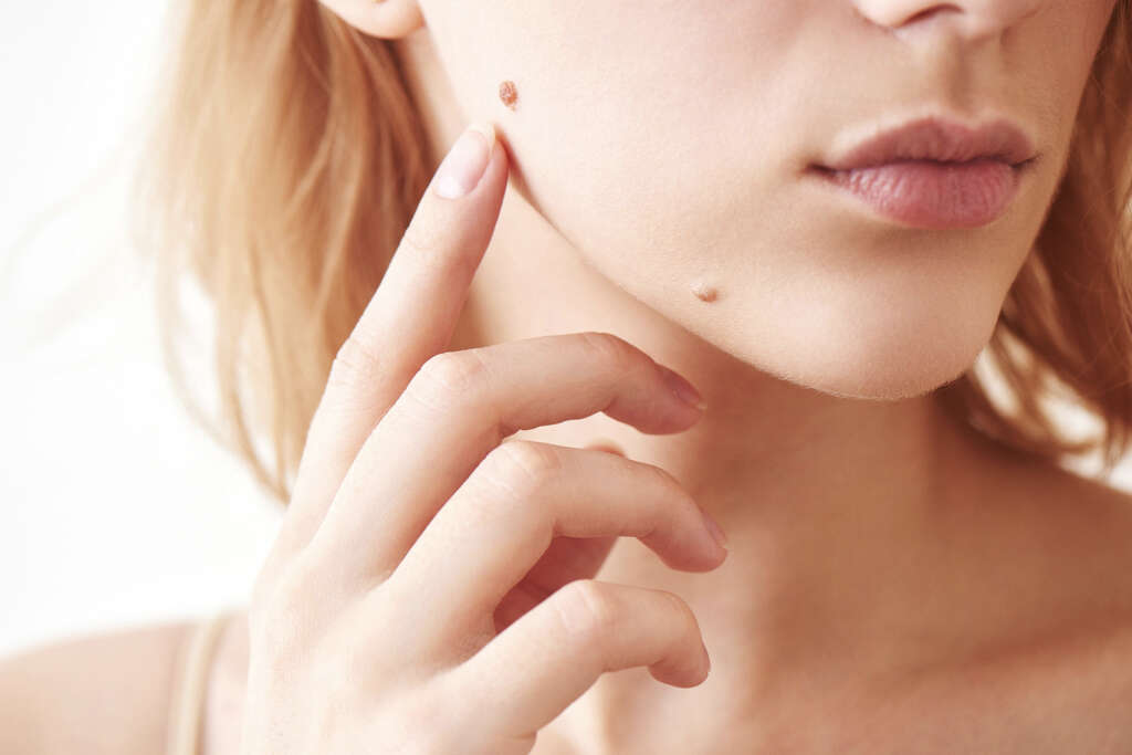
A mole or nevus (or moles/nevi if multiple) is a term used to describe a lesion on the skin that is generally brown or black. The term originates from the Latin word “nvus,” which translates to “birthmark.” A mole can be present at birth or acquired. It can appear anywhere on the skin and generally appears in early childhood or the first 25 years of life. By adulthood, it is normal to have 10 to 40 moles.
With time, the mole can change in appearance and may appear raised or have different colors. There may also be hair on the mole. While moles may develop with time, they may also disappear. Some other common terms often used to describe moles include birthmark and beauty mark.
1. Classification
Moles can be classified into hypermelanotic (increased melanin) and hypomelanotic (decreased melanin). In moles with increased melanin, they can broadly categorized into congenital (congenital melanocytic nevus, nevus of Ito, and nevus of Ota) and acquired (melanocytic nevus, atypical [dysplastic] nevus, Hori’s nevus, Becker’s nevus, Blue nevus, nevus spillus, spitz nevus, pigmented spindle cell nevus). In moles with decreased melanin, it includes nevus anemicus (acquired), nevus depigmentosus (congenital), connective tissue nevi (collagenoma, elastoma), epidermal nevi (eccrine nevus, apocrine nevus, nevus sebaceous, nevus comedonicus, and verrucous epidermal nevus), vascular nevi, and intramucosal nevi.

2. Composition
Melanocytic nevi are benign growths or hamartomas (tumor-like nodule) that are composed of melanocytes, which are cells that produce pigments. Melanocytes originate from the neural crest and migrate during development to sites such as the central nervous system, skin, ears, and eyes.
These cells produce melanin, a dark pigment mainly responsible for skin color. Moles at birth are thought to be an anomaly during development and can be considered as hamartomas or malformations. In contrast, acquired moles are thought of as benign neoplasms. When a melanocytic nevus becomes malignant, it is known as melanoma.
3. Mechanism
Melanocytes are melanin-producing cells that can be found in the basal layer of the skin (epidermis). Melanocytes usually exhibit contact inhibition and therefore pigment cells are generally not touching. However, some forms of stimulation, such as exposure to ultraviolet irradiation (sun), may increase the density of melanocytes in the skin.
Moles develop when the melanocytes that are in contact with each other proliferate and form collections of cells that are also known as nests. Moles usually form in the early childhood years. It is believed to be partly due to ultraviolet (sun) exposure, genetic factors, and blistering events.

4. Contributing Factors
Genetic factors have also been thought to play a role in the development of some melanocytic nevi. Some families with an autosomal dominant condition such as the familial atypical multiple mole and melanoma syndrome or dysplastic nevus syndrome have family members with a large number of large moles.
Moles have also been observed to develop after blistering events such as severe sunburns, genetic blistering diseases (epidermolysis bullosa), toxic epidermal necrolysis, and second degree thermal burns. Growth factors have also been thought to be contribute to the stimulation of melanocyte proliferation. As previously mentioned, moles present at birth are thought to be due to an anomaly during development where there is error in the migration of the melanocytes.

5. Causes
The exact cause of moles is still unknown. There are no established environmental or genetic influences that contribute to the development of congenital nevi. Researchers who studied 144 children in Naples concluded that the development of moles in early life is due to environmental factors, constitutional factors, anatomical location, and mole evolution.
The specific genetic factors are also unclear. However, experts believe that those with a large number of moles may have inherited an autosomal dominant trait. Individuals with dysplastic nevus syndrome and familial atypical multiple nevus have a higher risk of melanoma. Studies have observed that moles are common in those with poor sun tolerance.

6. Geographical Statistics
In the United States, acquired moles are so common that some experts do not consider it as an abnormality or defect. Although common, it represents an abnormal proliferation of cells. Most fair-skinned individuals will have at least several moles. It also occurs in dark-skinned individuals but at a lower prevalence.
Distribution differences of moles have also been observed with moles occurring more on the trunk in light skin, while in darker skin, moles tend to occur in the peripheral body parts. Globally, the prevalence of moles are similar to the United States. Individuals in Northern Europe such as Belgium, the Netherlands, and the United Kingdom have been observed to have large moles (equal or more than 1 centimeter in diameter) and have a large number of moles (more than 50).
7. Gender and Age Statistics
The appearance of moles are thought to be equal in both males and females. Judging the incidence and prevalence may be difficult as it is common and medical attention is usually only sought for cosmetic purposes. However, it has been observed that the moles may be responsive to sex hormones. This observation is supported by the changes in moles seen during pregnancy. In these cases, the moles commonly enlarge or darken.
Congenital moles are present at birth or soon after birth, while acquired moles are not present at birth. The incidence of moles increases throughout the first 30 years and peaks in the 4th to 5th decades of life. After that, the incidence decreases as the mole slowly involutes and becomes inconspicuous in advanced elder years.
8. Mole Removal
When the risk of malignancy is increased, histopathological examination and other tests may be necessary. Removal of the mole may be indicated when there is potential malignancy or for cosmetic reasons. Tangential or shave excision can be performed for cosmetic removal, punch excision for small lesions, and a complete excision with sutured closure for large lesions (more than 1 cm in diameter).
The excised mole can be sent to a pathologist so an accurate diagnosis can be made to confirm if the mole is malignant. Referral to a dermatologist should be made if there are any doubts regarding the diagnosis and management of the mole. This is especially true if melanoma is suspected.
9. Prognosis
The prognosis or outcome of having a single mole is excellent as these lesions are considered benign growths that have no potential to turn malignant (unless evolution occurs). Those with multiple moles or large moles may have a higher risk of melanoma with the risk increasing with higher number and bigger sizes.
Moles are biologically stable; however, they can also be associated with melanoma (skin cancer). The true rate of transformation of a mole to melanoma is unknown with some data suggesting that about 40% of melanoma cases have a precursor mole. The risk of melanoma is higher in congenital moles (especially in large moles). Some studies have suggested that melanoma may occur in up to 5% of giant congenital moles.

10. Patient Education
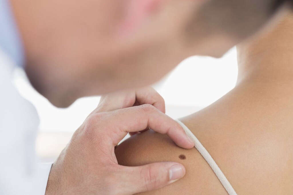
Patients should be made aware that moles are a common anomaly that are benign and do not generally cause any issues. However, they should be taught self-examination of their moles (especially those with a higher risk-large congenital moles). Use the ABCDE approach, where patients assess the asymmetry, border irregularity, color, diameter, and evolution of the mole.
Some add an “F” for funny looking, which suggests that the appearance of the mole has changed. Other things to look out for include raised borders, itchiness, and bleeding. Those with these changes should seek medical attention to determine the potential of malignant transformation. There are many resources and information available that illustrate these changes from various organizations such as the American Academy of Dermatology. The importance of sun protection should be emphasized for both children and adults.


