What Are Hemangiomas?

 Article Sources
- 1. Freelove, DDS, Cameron. 'Hemangioma.' Center for Craniofacial Molecular Biology, USC, Herman Ostrow School of Dentistry of USC, Nov. 2017, ccmb.usc.edu/files/2017/11/StudentLectures7-11.pdf.
- 2. Mulliken, MD, John B., and Odile Enjolras. 'Congenital Hemangiomas and Infantile Hemangioma: Missing Links.' Molecular and Cellular Mechanisms of Vascular Anomalies, Harvard School of Dental Medicine, June 2004, vascularanomalies.hsdm.harvard.edu/Publications/Mulliken8.pdf.
- 3. Tafti, Dawood, and Nathan D. Cecava. 'Spinal Hemangioma.' National Center for Biotechnology Information, U.S. National Library of Medicine, 8 Aug. 2020, www.ncbi.nlm.nih.gov/books/NBK532997.
- 4. 'Hemangiopericytoma.' Genetic and Rare Diseases Information Center, U.S. Department of Health and Human Services, rarediseases.info.nih.gov/diseases/2627/hemangiopericytoma.
- 5. 'Hemangioblastoma.' NIH GARD, U.S. Department of Health & Human Services, rarediseases.info.nih.gov/diseases/8232/hemangioblastoma.
- 6. John H. Greinwald Jr, MD. 'An Update on the Treatment of Hemangiomas in Children With Interferon Alfa-2a.' Archives of Otolaryngology–Head & Neck Surgery, JAMA Network, 1 Jan. 1999, jamanetwork.com/journals/jamaotolaryngology/fullarticle/508995.
Vascular anomalies are often blood vessel growths. Most people refer to visible vascular anomalies as birthmarks. Doctors categorize these anomalies as either vascular tumors or vascular malformations. The most well-known type of vascular tumor is the hemangioma, a tumor composed of blood vessels.
Most people are familiar with the form of hemangiomas that appear on an infant soon after birth. Approximately 5 to 10 percent of infants have one.1Freelove, DDS, Cameron. ‘Hemangioma.’ Center for Craniofacial Molecular Biology, USC, Herman Ostrow School of Dentistry of USC, Nov. 2017, ccmb.usc.edu/files/2017/11/StudentLectures7-11.pdf. Hemangiomas are benign, which means non-cancerous, and they may appear anywhere on the body, including the skin and organs. Though they aren’t cancerous, sometimes their location can cause dangerous complications.
1. Hemangiomas
Hemangiomas in infants may present as a mass of blood vessels on the surface of the skin, in the fat beneath the skin, or as a combination of both. Most of them get larger and puffier during the first year. Then they eventually shrivel and fade away. However, when their placement impacts breathing or vision, medical intervention may be required.
Cherry hemangiomas are found on the skin of adults as tiny red-purple bumps. They usually require no treatment. Internal hemangiomas are also benign. But treatment may be necessary if their location causes pain, bleeding, or other dangerous symptoms.
2. Infantile Hemangiomas
Parents may refer to these common hemangiomas as birthmarks, although they form after birth. Typically, they first appear as a light red mark a few weeks to a few months after birth. But they grow quickly over the first year of life. Girls experience these masses three to five times more often than boys.
Infantile hemangiomas generally shrink away by the child’s fifth birthday. Sometimes, scar tissue remains. Because they normally fade away naturally, infantile hemangiomas usually don’t require treatment. Exceptions may occur when complications arise.

3. Congenital Hemangiomas
Congenital hemangiomas form before birth, in the uterus. They don’t experience rapid growth like infantile hemangiomas. Congenital hemangiomas are more common among Caucasian infants than African American or Asian babies. Males and females experience these tumors about equally.
Doctors categorize these masses as rapidly involuting congenital hemangiomas (RICH) or noninvoluting congenital hemangioma (NICH). RICH usually appear on the arms, legs, neck or head, and they shrink soon after birth. Sometimes surgery is required. NICH are very rare. They don’t shrink on their own and may need to be surgically removed.2Mulliken, MD, John B., and Odile Enjolras. ‘Congenital Hemangiomas and Infantile Hemangioma: Missing Links.’ Molecular and Cellular Mechanisms of Vascular Anomalies, Harvard School of Dental Medicine, June 2004, vascularanomalies.hsdm.harvard.edu/Publications/Mulliken8.pdf.
4. Spinal Hemangiomas
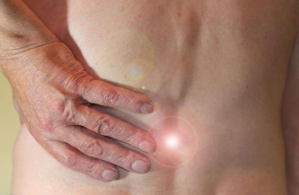
Spinal hemangiomas are a typical form of benign spinal tumor. Approximately 11 percent of the population may have this type of tumor.3Tafti, Dawood, and Nathan D. Cecava. ‘Spinal Hemangioma.’ National Center for Biotechnology Information, U.S. National Library of Medicine, 8 Aug. 2020, www.ncbi.nlm.nih.gov/books/NBK532997. But these growths often don’t cause symptoms, so people are unaware they have them. For those who do experience symptoms, back pain is the most typical symptom.3Tafti, Dawood, and Nathan D. Cecava. ‘Spinal Hemangioma.’ National Center for Biotechnology Information, U.S. National Library of Medicine, 8 Aug. 2020, www.ncbi.nlm.nih.gov/books/NBK532997.
Men and women between their 40s and 60s are the population segment most likely to have spinal hemangiomas. Women appear to experience symptoms more often than men. Treatment is only necessary if there’s significant pain or neurological issues.
5. Internal Organ Hemangiomas
Most people never know they have an internal hemangioma since symptoms are rare. While they can form on or within any organ, they’re most common on the intestines, liver, and brain. Liver hemangiomas bigger than 1.6 inches may produce symptoms like fullness, nausea and weight loss.
Hemangioblastomas and hemangiopericytomas are the two types of hemangiomas that may form in the brain. They’re both benign in most cases. Hemangioblastomas only represent 2 percent of all brain tumors. Hemangiopericytomas are even rarer and can be either non-cancerous or malignant (cancerous).4‘Hemangiopericytoma.’ Genetic and Rare Diseases Information Center, U.S. Department of Health and Human Services, rarediseases.info.nih.gov/diseases/2627/hemangiopericytoma. They usually grow in the meninges that cover the brain.5‘Hemangioblastoma.’ NIH GARD, U.S. Department of Health & Human Services, rarediseases.info.nih.gov/diseases/8232/hemangioblastoma.
6. Intramuscular Hemangiomas
Vascular tumors within the muscle are called intramuscular hemangiomas. They’re a rare form of hemangioma. These masses are often painful and difficult to diagnose due to location and rarity. Intramuscular hemangiomas can affect any muscle. Activity can cause swelling and more discomfort.
Young adults are most susceptible to these soft tissue tumors, and women are slightly more likely to develop these growths. Trauma appears to be the most common cause. The thigh muscle is a typical location for this type of hemangioma.
7. Common Locations
Hemangiomas can form anywhere. That includes the surface of the skin or just beneath it on the face, neck, head, back, torso, and limbs. They can also grow on internal organs, muscle, and bone.
Skin formation is most common. Infantile hemangiomas are typically this type. In cavernous hemangioma, loosely packed, widened, larger blood vessels form a mass or tumor. They form in the skin and inside the body. Sizes vary. Compound hemangiomas are a combination of the cavernous and capillary forms.
8. Complications
Among infants, the most prevalent complications occur when the hemangioma is in a dangerous location. If it’s close to the eye, vision issues can occur. In the infant’s neck, there may be difficulty breathing. Bleeding, ulcers, infections, and deformity are other potential problems.
Other types of hemangiomas can present risks. For example, a hemangioblastoma can cause neurological issues as it grows and puts pressure on the brain. Other internal hemangiomas may present problems and complications when they become symptomatic. Medical diagnoses, evaluation, and treatment may be necessary.

9. Causes
There is no single known cause for hemangiomas. But statistics show a correlation between premature birth and infantile hemangiomas. These benign tumors are also more common in low birth weight Caucasian babies. Additionally, some experts suspect a genetic factor in some cases, and many families experience more hemangiomas among their members than the average occurrence.
Cherry hemangiomas have been linked to pregnancy, climate, some medical conditions, and chemical exposure. They also usually become more common with age, especially after year 30.
10. Treatment
Treatment isn’t usually necessary for most hemangiomas. That’s because they’re generally asymptomatic and go away on their own. But when complications occur, or the risk of complications is high, medical intervention may be necessary.
Some common treatment options include medicated gels, corticosteroids, laser treatments, radiation therapy, and steroids. Propranolol is a beta-blocker sometimes used to shrink infantile hemangiomas. Interferon alfa-2a therapy can be used in severe forms of hemangiomas.6John H. Greinwald Jr, MD. ‘An Update on the Treatment of Hemangiomas in Children With Interferon Alfa-2a.’ Archives of Otolaryngology–Head & Neck Surgery, JAMA Network, 1 Jan. 1999, jamanetwork.com/journals/jamaotolaryngology/fullarticle/508995. Medical professionals may treat cherry hemangiomas with electrocauterization, laser surgery, or cryosurgery. However, hemangiomas can recur after surgery.