10 Hemangioma Symptoms
A hemangioma refers to a benign tumor that occurs due to the proliferation of vascular endothelial cells. The commonest type of hemangioma is known as the infantile hemangioma, which is commonly seen in infancy. Also known as strawberry marks, hemangiomas are often noticed at birth or during the first few weeks of life.
A hemangioma can occur on any part of the body. However, the commonest sites are the face, chest, scalp, and back.
Since hemangiomas usually fade with time, treatment is usually unnecessary as therapeutic options have potential side effects. However, hemangiomas that cause symptoms can be treated using corticosteroids, beta blockers, and laser surgery.

Symptom #1: Macules and Papules
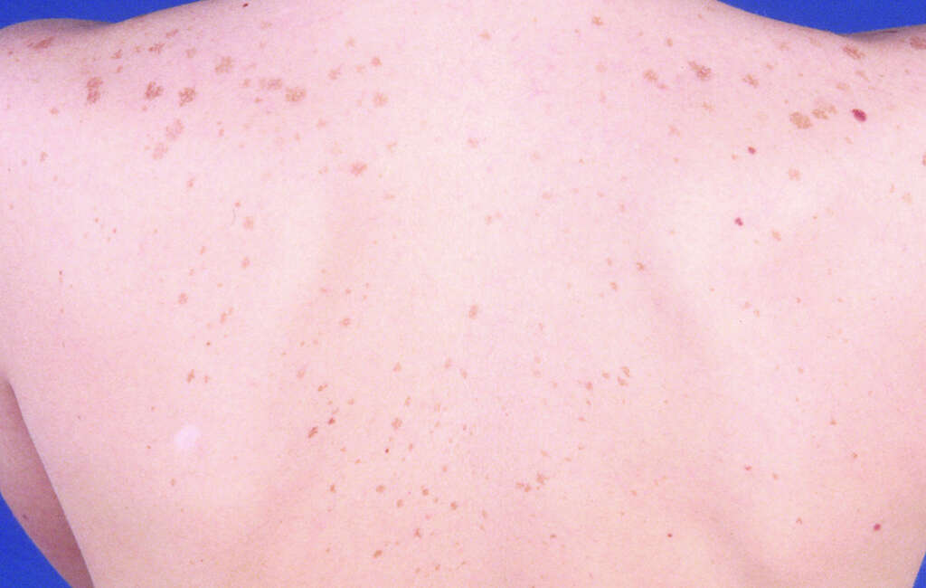
Macules are defined as small circumscribed spots on the skin that are not depressed or elevated. Macules are usually small and can be appreciated through visual inspection. Papules are similar to macules but are elevated and can be felt as a bump on the skin.
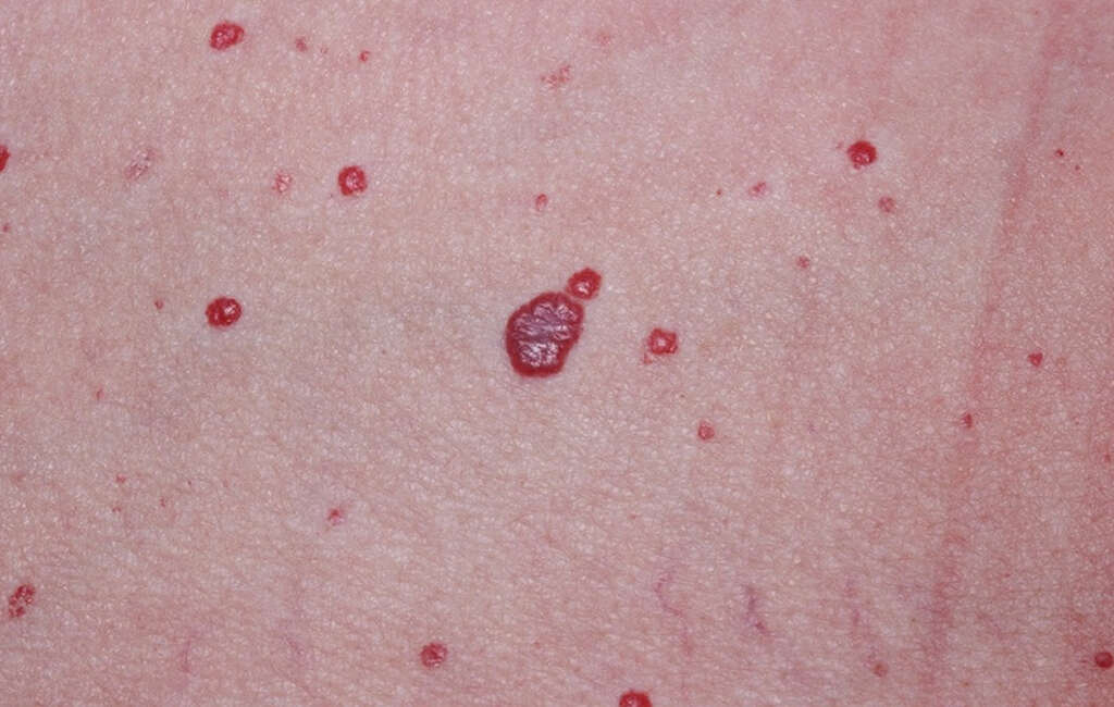
Cherry hemangiomas or angiomas are also known as senile angiomas or Campbell De Morgan spots. These hemangiomas are usually small and cherry red in color. They are harmless and occur due to the abnormal proliferation of blood vessels. It is the commonest type of angioma and is typically seen in almost all adults over the age of 30 years old.
Symptom #2: Bleeding
Bleeding occurs when there is blood escaping from the circulatory system. It can occur internally or externally. Although hemangiomas are usually harmless and do not cause symptoms, bleeding can occur if there is injury to the hemangioma. It can also become inflamed or thrombosed.
Those with profuse bleeding that does not stop should seek medical attention. Hemangiomas that stop bleeding should be cleaned and proper wound care is important to prevent the wound from getting infected.
Symptom #3: Ulceration
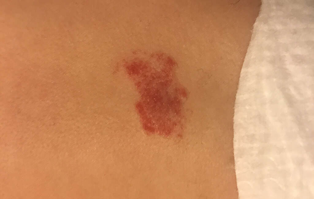
Ulceration can also occur and has been observed in 10 to 15 percent of infantile hemangiomas. Although the cause of ulceration is unclear, it has been thought to be due to the action of some cytokines or because of the outstripped blood supply for the skin overlying the hemangioma.
Ulceration is usually seen only in hemangiomas that are tense and rapidly proliferating. Ulceration is also more commonly observed in hemangiomas that occur on the chest, lip, or anogenital region. However, it is important to note that it can occur at any site. Ulcerations are very painful and lead to the formation of a scar once healed. In early stages before ulceration actually occurs, a white discoloration can be seen. This mimics involution.

Symptom #4: Infection
Infection occurs when the body has been invaded by disease-causing pathogens. These pathogens then multiply causing the immune system to attempt to fight it off. Infections can be caused by bacteria, viruses, parasites, prions, and fungi. Inflammation usually occurs when there is an infection. An infected hemangioma can become painful, swollen, red, and warm.
Bleeding or ulcerated hemangiomas should be treated to prevent infection. The treatment for ulcerated hemangiomas includes the use of topical anesthetics, bio-occlusive dressings, topical timolol, pulsed-dye laser surgery, and oral propranolol. Depending on the organism causing the infection, different medications may also be administered.
Symptom #5: Visual Loss
Visual impairment occurs when there is decreased ability to see which is not fixable through the use of glasses. Visual impairment or loss is important as it causes difficulty with everyday activities. The commonest causes of visual impairment are cataracts, glaucoma, and uncorrected refractive errors. Other conditions that can cause visual issues include diabetic retinopathy, age-related macular degeneration, and childhood blindness.
Visual loss can occur when a hemangioma occurs on the eyelids or periorbital tissues. There may be a gradual inability to open the affected eye as there is progressive involvement of the eyelid.

Symptom #6: Exophthalmos or Proptosis
Exophthalmos or proptosis refers to the bulging out of the eyes anteriorly or in the forward direction. It can be bilateral or unilateral. Bilateral exophthalmos is usually seen in Grave’s disease while unilateral cases are often due to a growth occurring behind the eye.
Exophthalmos is significant as it can result in complete or partial dislocation from the orbit in severe cases. It can also occur due to trauma. Left untreated, the eyelids are unable to close over the eyes during sleep and blinking. This results in corneal dryness and damage. Another possible complication is the compression of the ophthalmic artery or optic nerve, resulting in blindness. In cases where the hemangioma extends posteriorly behind the eye, it can result in proptosis.

Symptom #7: Amblyopia
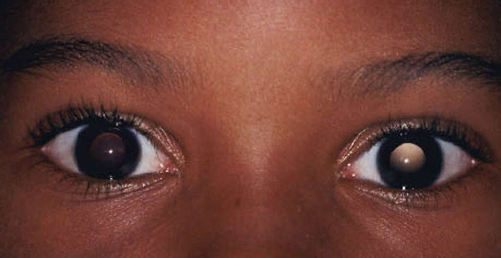
Amblyopia or lazy eye occurs when the eye and brain do not work well together, resulting in decreased vision despite having normal eyes. It is the commonest cause of decreased vision among children and adolescents. Amblyopia can be caused by any condition that interferes with the focus of the eyes in early childhood. Conditions that cause amblyopia include poor alignment of the eyes, irregular shaped eyes, and clouding of the lens.
In cases of hemangioma, amblyopia can occur in a visual obstruction that has been left untreated. Even after the removal of the hemangioma, vision will not be restored immediately. If untreated, amblyopia persists into adulthood.

Symptom #8: Anisometropia
Anisometropia refers to two eyes that have different refractive power. Each eye can be farsighted (hyperopia), nearsighted (myopia), or have antimetropia (combination of hyperopia and myopia). Anisometropia can be defined as two eyes that have a difference in power of two diopters or more.
In some cases, the brain will suppress the central vision from one of the eyes, resulting in amblyopia (if occurring during the first 10 years of life due to the development of the visual cortex). The management of anisometropia includes the use of corrective glasses, contact lenses, or refractive surgery. In hemangioma, the involvement of the eye can result in anisometropia.
Symptom #9: Thrombocytopenia
Thrombocytopenia is a term describing abnormally low levels of platelets. It is usually asymptomatic and is only noticed when a full blood count is performed. In some cases, patients may experience bleeding gums, nosebleeds, easy bruising, and heavy periods. Petechiae may also appear. Some patients may also complain of fatigue, malaise, and general weakness.
In some syndromes such as the Kasabach–Merritt syndrome, the presence of giant hepatic hemangiomas has been associated with intravascular coagulation and thrombocytopenia. Patients are usually younger than 1 year old and are male. In this case, the hemangioma should be eradicated along with gaining control of the patient’s coagulopathy.
Symptom #10: Congestive Heart Failure
Congestive heart failure occurs when the amount of blood pumped by the heart is more than normal. This increased workload results in the heart being unable to keep up. This can be seen in patients with chronic severe anemia, kidney diseases, hyperthyroidism, beriberi, multiple myeloma, cirrhosis, and Paget’s disease.
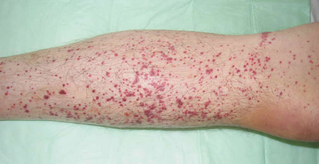
Patients with heart failure may experience shortness of breath, pedal edema, excessive tiredness, and coughing. Congestive heart failure may occur in patients who have multiple hemangiomas. Those with diffuse neonatal hemangiomatosis will have multiple cutaneous and visceral hemangioma. This results in congestive heart failure due to the increased vascular volume.


