What Is Charcot Foot?

 Article Sources
- 1. Kaynak, Gökhan et al. “An overview of the Charcot foot pathophysiology.” Diabetic Foot and Ankle vol. 4 (2013): 10.3402/dfa.v4i0.21117.
- 2. Roberts, Lee C. et al. “The Charcot Foot in Diabetes.” Diabetes Care, vol. 34(9) (2011): 2123-2129.https://doi.org/10.2337/dc11-0844
- 3. Shah, Mrugeshkumar et al. “Charcot Arthropathy.” Medscape Drugs and Diseases (2020): https://emedicine.medscape.com/article/1234293-overview
Also called Charcot arthropathy or neuropathic joint, Charcot foot is a potentially debilitating deformity of the foot in which the plantar arch collapses and bows outward into what is known as a rocker-foot. Patients with this condition walk with difficulty but may experience relatively little pain due to pre-existing numbness in the feet.3Shah, Mrugeshkumar et al. “Charcot Arthropathy.” Medscape Drugs and Diseases (2020): https://emedicine.medscape.com/article/1234293-overview
The condition gets its name from the French neurologist, Jean-Martin Charcot (1825-1893), who first described it.1Kaynak, Gökhan et al. “An overview of the Charcot foot pathophysiology.” Diabetic Foot and Ankle vol. 4 (2013): 10.3402/dfa.v4i0.21117. It is most often associated with diabetes, but anyone experiencing nerve damage to the extremities is potentially at risk. Here are 10 important facts to know about Charcot foot.
1. Causes
Once thought to be caused by syphilis, Charcot foot is most widespread among diabetics in the United States3Shah, Mrugeshkumar et al. “Charcot Arthropathy.” Medscape Drugs and Diseases (2020): https://emedicine.medscape.com/article/1234293-overview and people with leprosy in regions with large indigenous populations.1Kaynak, Gökhan et al. “An overview of the Charcot foot pathophysiology.” Diabetic Foot and Ankle vol. 4 (2013): 10.3402/dfa.v4i0.21117. However, a variety of conditions resulting in neuropathy also can cause the condition, including nerve injuries, infections, steroid use, and heavy metal poisoning.1Kaynak, Gökhan et al. “An overview of the Charcot foot pathophysiology.” Diabetic Foot and Ankle vol. 4 (2013): 10.3402/dfa.v4i0.21117.
Because those with neuropathy have a diminished sense of pain in the feet, they are vulnerable to traumatic injuries in that area. For example, they may sprain an ankle or step on a nail without realizing it. Tissue damage can then lead to severe inflammation,2Roberts, Lee C. et al. “The Charcot Foot in Diabetes.” Diabetes Care, vol. 34(9) (2011): 2123-2129.https://doi.org/10.2337/dc11-0844 and the inflammation to loss of bone tissue and eventual fractures.3Shah, Mrugeshkumar et al. “Charcot Arthropathy.” Medscape Drugs and Diseases (2020): https://emedicine.medscape.com/article/1234293-overview
2. Anatomical Classification
Charcot foot is so-named because it most often affects the foot and ankle. Specifically, it is most likely to occur in the tarsometatarsal joint and the cuneonavicular, talonavicular, and calcaneocuboid articulations. However, it can develop in any joint.3Shah, Mrugeshkumar et al. “Charcot Arthropathy.” Medscape Drugs and Diseases (2020): https://emedicine.medscape.com/article/1234293-overview
At least three classification systems exist to describe the pattern of incidence in the body of Charcot syndrome and to classify its severity. Most doctors refer to the Saunders and Mrdjencovich system of five foot and ankle regions to both describe the condition and predict its progression.3Shah, Mrugeshkumar et al. “Charcot Arthropathy.” Medscape Drugs and Diseases (2020): https://emedicine.medscape.com/article/1234293-overview
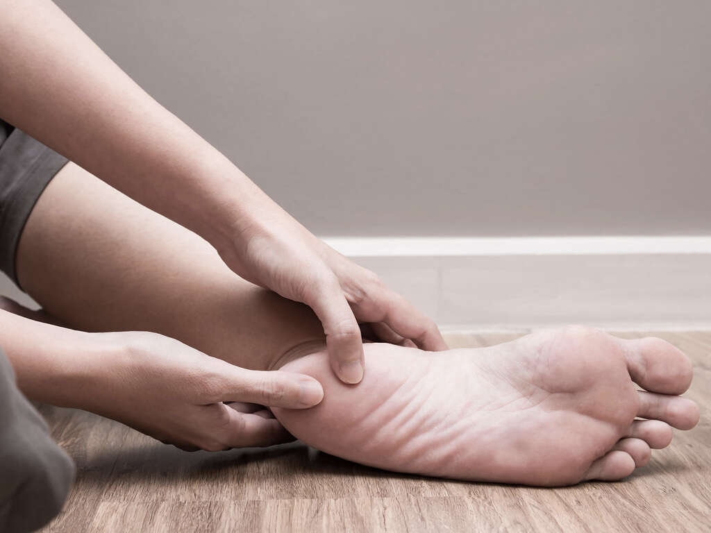
3. Symptoms
The primary symptom is a “rocker-bottom” foot in which the arch has collapsed, creating a convex sole on which it is difficult to walk.2Roberts, Lee C. et al. “The Charcot Foot in Diabetes.” Diabetes Care, vol. 34(9) (2011): 2123-2129.https://doi.org/10.2337/dc11-0844 This deformity can range from mild to debilitating.3Shah, Mrugeshkumar et al. “Charcot Arthropathy.” Medscape Drugs and Diseases (2020): https://emedicine.medscape.com/article/1234293-overview However, most patients report feeling much less pain than would be expected, due to being insensate in the affected area.3Shah, Mrugeshkumar et al. “Charcot Arthropathy.” Medscape Drugs and Diseases (2020): https://emedicine.medscape.com/article/1234293-overview
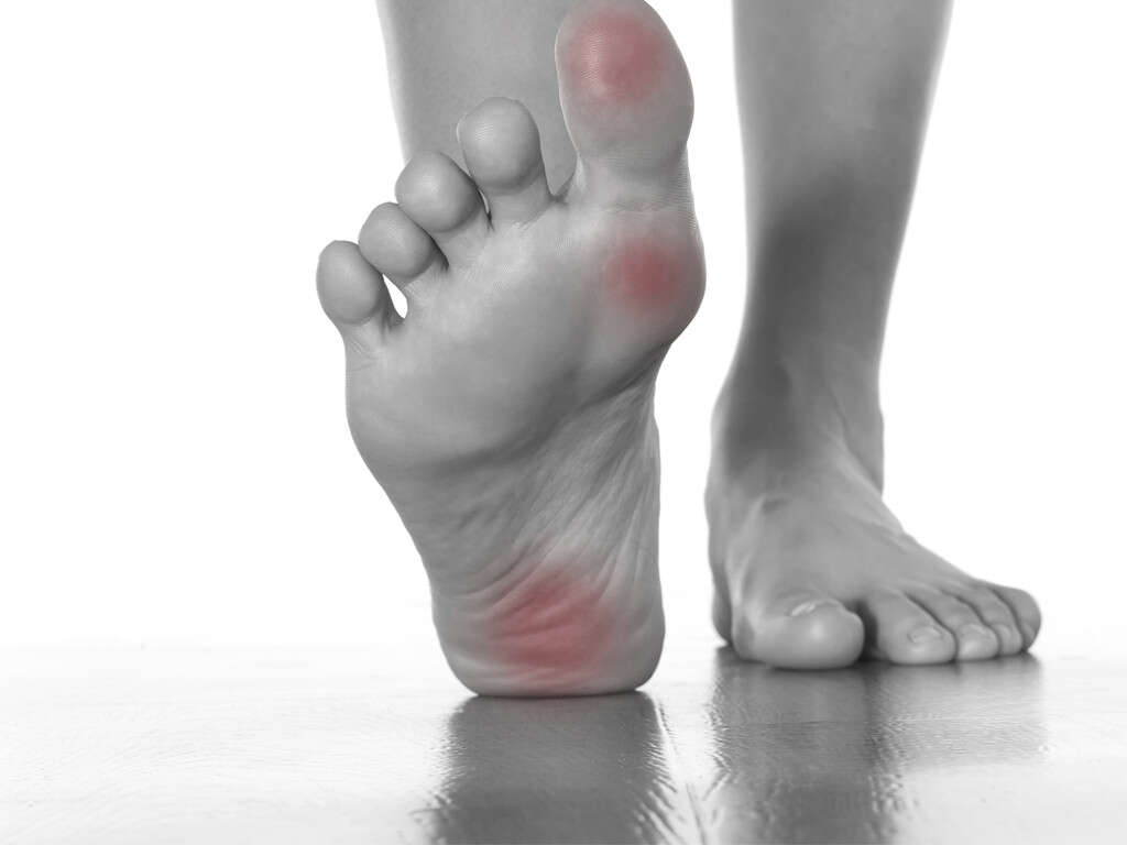
Signs of inflammation that may precede bone deformities are swelling, redness, and warmth in one foot.2Roberts, Lee C. et al. “The Charcot Foot in Diabetes.” Diabetes Care, vol. 34(9) (2011): 2123-2129.https://doi.org/10.2337/dc11-0844 Other symptoms include joint effusion, joint instability, and bone resorption. Foot ulcers and callused areas are common on the skin of the foot, especially covering places where bones protrude.3Shah, Mrugeshkumar et al. “Charcot Arthropathy.” Medscape Drugs and Diseases (2020): https://emedicine.medscape.com/article/1234293-overview
4. Occurrence
Various sources report Charcot foot to occur in 0.15%3Shah, Mrugeshkumar et al. “Charcot Arthropathy.” Medscape Drugs and Diseases (2020): https://emedicine.medscape.com/article/1234293-overview to up to 13% of diabetics and in 1% to 29% of patients with neuropathy.1Kaynak, Gökhan et al. “An overview of the Charcot foot pathophysiology.” Diabetic Foot and Ankle vol. 4 (2013): 10.3402/dfa.v4i0.21117. One author reports a delay in diagnosis in 25% of patients, resulting in probable underestimates of occurrence. Overall, cases are increasing1Kaynak, Gökhan et al. “An overview of the Charcot foot pathophysiology.” Diabetic Foot and Ankle vol. 4 (2013): 10.3402/dfa.v4i0.21117..
Some analyses detect no disparity in the sexes, while others have found it to occur three times more frequently in men than women. 10% of patients experience symptoms in both feet, and less than 5% have a recurrence of the condition after treatment.3Shah, Mrugeshkumar et al. “Charcot Arthropathy.” Medscape Drugs and Diseases (2020): https://emedicine.medscape.com/article/1234293-overview Occurrence worldwide is uncertain.
5. Prognosis
After treatment, the foot and ankle require 38 to 113 days to heal, depending on the specific area damaged. The forefoot is the area quickest to recover, and the heel the slowest. Full recovery is expected in one to two years. However, the patient must take special care of the foot for the rest of his or her life.3Shah, Mrugeshkumar et al. “Charcot Arthropathy.” Medscape Drugs and Diseases (2020): https://emedicine.medscape.com/article/1234293-overview
Diabetics generally take longer to recover from injuries, due in part to reduced blood circulation. The more serious the injury, the longer the recovery and the greater chance of permanent damage.3Shah, Mrugeshkumar et al. “Charcot Arthropathy.” Medscape Drugs and Diseases (2020): https://emedicine.medscape.com/article/1234293-overview
6. Diagnosis
Doctors sometimes misdiagnose early signs of Charcot foot as gout, thrombosis, or cellulitis, due to the similarity in inflammation among the conditions. Many will X-ray the foot or conduct an MRI to provide an image of the bones.2Roberts, Lee C. et al. “The Charcot Foot in Diabetes.” Diabetes Care, vol. 34(9) (2011): 2123-2129.https://doi.org/10.2337/dc11-0844 Other bone tests sometimes performed are bone probing, synovial biopsy, and joint aspiration.3Shah, Mrugeshkumar et al. “Charcot Arthropathy.” Medscape Drugs and Diseases (2020): https://emedicine.medscape.com/article/1234293-overview
Laboratory tests include a white blood cell count and an erythrocyte sedimentation rate, both of which are used to rule out osteomyelitis as causing the bone deformities. A basic metabolic profile is another blood test often performed to test for the contributing factors of diabetes, bone diseases, cancer, infections, and alcoholism.3Shah, Mrugeshkumar et al. “Charcot Arthropathy.” Medscape Drugs and Diseases (2020): https://emedicine.medscape.com/article/1234293-overview
7. Treatment
Patients are required to wear a cast on the injured foot for an average of 12.5 to 18.5 weeks. The cast serves to restrict movement, alleviate pressure, and prevent further injury during healing. When the cast is no longer necessary, patients must wear a brace and special shoes for up to two years.3Shah, Mrugeshkumar et al. “Charcot Arthropathy.” Medscape Drugs and Diseases (2020): https://emedicine.medscape.com/article/1234293-overview
Surgery is necessary in cases of extreme deformity, when the foot will not fit into a cast or brace, or when other methods have proven unsuccessful. Surgeons work to even out the sole of the foot so that it can fit into a shoe and allow the patient to walk. Less than one-fourth of patients require surgery.3Shah, Mrugeshkumar et al. “Charcot Arthropathy.” Medscape Drugs and Diseases (2020): https://emedicine.medscape.com/article/1234293-overview
8. Complications
Besides rocker-foot, other deformities can occur. For example, the entire foot can collapse into a clubfoot and the Lisfranc joint complex may suffer fractures and dislocations. Furthermore, ligaments can ossify, intra-articular and extra-articular exostoses form, and osteomyelitis develop.3Shah, Mrugeshkumar et al. “Charcot Arthropathy.” Medscape Drugs and Diseases (2020): https://emedicine.medscape.com/article/1234293-overview
The most extreme complication from Charcot foot is amputation of the extremity due to an infected skin ulcer. This situation can occur when a fracture is not treated. As the patient continues to walk on the injured bone, ulcers can form over the bony protrusions, which then run a risk of infection.3Shah, Mrugeshkumar et al. “Charcot Arthropathy.” Medscape Drugs and Diseases (2020): https://emedicine.medscape.com/article/1234293-overview
9. Potential Treatments
Medications with the potential to help arrest bone deterioration are bisphosphonates. Theoretically, these compounds should not only stop bones from breaking down but also reduce inflammation and help the bone to heal itself. So far, they have not been used widely.3Shah, Mrugeshkumar et al. “Charcot Arthropathy.” Medscape Drugs and Diseases (2020): https://emedicine.medscape.com/article/1234293-overview
Two non-invasive therapies under study are low-intensity ultrasound and magnetic field therapy. The former has been shown to improve strength and decrease the time it takes for a bone to heal. The latter has some evidence of increased healing time, but no other proof of benefits.3Shah, Mrugeshkumar et al. “Charcot Arthropathy.” Medscape Drugs and Diseases (2020): https://emedicine.medscape.com/article/1234293-overview
10. Management
After treatment with a cast, patients must take special care of their feet for at least the next one to two years. Several kinds of protective braces are available, including a patellar tendon-bearing brace, a double metal ankle-foot orthosis, a shoe containing a modified ankle-foot orthosis, or a Charcot restraint orthotic walker.3Shah, Mrugeshkumar et al. “Charcot Arthropathy.” Medscape Drugs and Diseases (2020): https://emedicine.medscape.com/article/1234293-overview
Special shoes are available to support healing for up to two years after cast removal. This footwear includes metal- or plastic-shanked shoes with firm soles and extra room inside the shoe. Some have a specialized sole to accommodate rocker-bottom feet and relieve pressure on ulcers.3Shah, Mrugeshkumar et al. “Charcot Arthropathy.” Medscape Drugs and Diseases (2020): https://emedicine.medscape.com/article/1234293-overview